- English
- Chinese
- French
- German
- Portuguese
- Spanish
- Russian
- Japanese
- Korean
- Arabic
- Irish
- Greek
- Turkish
- Italian
- Danish
- Romanian
- Indonesian
- Czech
- Afrikaans
- Swedish
- Polish
- Basque
- Catalan
- Esperanto
- Hindi
- Lao
- Albanian
- Amharic
- Armenian
- Azerbaijani
- Belarusian
- Bengali
- Bosnian
- Bulgarian
- Cebuano
- Chichewa
- Corsican
- Croatian
- Dutch
- Estonian
- Filipino
- Finnish
- Frisian
- Galician
- Georgian
- Gujarati
- Hausa
- Hawaiian
- Hebrew
- Hmong
- Hungarian
- Icelandic
- Igbo
- Javanese
- Kannada
- Kazakh
- Khmer
- Kurdish
- Kyrgyz
- Latin
- Latvian
- Lithuanian
- Luxembou..
- Macedonian
- Malagasy
- Malay
- Malayalam
- Maltese
- Maori
- Marathi
- Mongolian
- Burmese
- Nepali
- Norwegian
- Pashto
- Persian
- Punjabi
- Serbian
- Sesotho
- Sinhala
- Slovak
- Slovenian
- Somali
- Samoan
- Scots Gaelic
- Shona
- Sindhi
- Sundanese
- Swahili
- Tajik
- Tamil
- Telugu
- Thai
- Ukrainian
- Urdu
- Uzbek
- Vietnamese
- Welsh
- Xhosa
- Yiddish
- Yoruba
- Zulu
- Kinyarwanda
- Tatar
- Oriya
- Turkmen
- Uyghur

Liver Cancer Explained: Symptoms, Causes & Expert Guide
2026-06-19
- What Is Cancer in Liver and How Does It Develop?
- Key Symptoms and Warning Signs of Liver Cancer
- Primary Causes and Risk Factors
- Diagnostic Methods and Screening Protocols
- Staging Systems and Prognosis
- Treatment Options and Management Strategies
- Prevention and Risk Reduction
- Comparison of Diagnostic Approaches
- Frequently Asked Questions (FAQ)
- Conclusion and Next Steps
Cancer in liver tissues, medically known as hepatocellular carcinoma or intrahepatic cholangiocarcinoma, occurs when healthy cells mutate and grow uncontrollably. This condition often develops in individuals with chronic liver disease, such as cirrhosis or hepatitis infections. Early detection is critical because symptoms frequently remain hidden until the disease advances. Understanding the causes, recognizing early warning signs, and knowing modern diagnostic methods are essential steps for effective management and improved survival rates.
What Is Cancer in Liver and How Does It Develop?
Cancer in liver refers to malignant growths originating within the liver itself, distinct from cancers that spread to the liver from other organs. The liver performs over 500 vital functions, including detoxification, protein synthesis, and bile production. When cellular DNA damages accumulate without repair, normal regulation fails, leading to tumor formation.
The development process typically begins with chronic inflammation. Over years, repeated injury causes scar tissue formation, known as fibrosis, which can progress to cirrhosis. In this compromised environment, genetic mutations become more likely. These mutations drive cells to divide rapidly, ignoring signals to stop or die.
There are several primary types of liver cancer. Hepatocellular carcinoma (HCC) is the most common form, accounting for the majority of cases. It starts in hepatocytes, the main liver cell type. Another type, cholangiocarcinoma, begins in the bile ducts. Less common forms include hepatoblastoma, mostly affecting children, and angiosarcoma, which arises in blood vessels.
The Role of Chronic Liver Disease
Chronic liver disease acts as the primary catalyst for most liver cancer cases. Conditions like chronic hepatitis B or C create a persistent inflammatory state. The immune system constantly fights the virus, causing collateral damage to liver tissue. This cycle of damage and regeneration increases the risk of errors during cell division.
Cirrhosis represents the end-stage of many liver diseases. In cirrhotic livers, normal architecture is replaced by nodules of regenerating cells surrounded by scar tissue. These nodules are prone to malignant transformation. Industry experts普遍认为 that monitoring patients with cirrhosis is the most effective strategy for early detection.
Key Symptoms and Warning Signs of Liver Cancer
Identifying cancer in liver early is challenging because the organ has significant functional reserve. Symptoms often do not appear until the tumor is large or liver function is severely impaired. Recognizing subtle changes can lead to earlier diagnosis and better outcomes.
Early-stage symptoms are often vague and easily mistaken for less serious conditions. Patients might experience unexplained fatigue, mild abdominal discomfort, or a general sense of feeling unwell. As the disease progresses, specific signs become more apparent.
- Unintended weight loss: Rapid loss of appetite and body mass without dieting.
- Abdominal pain: Discomfort or swelling in the upper right abdomen, near the rib cage.
- Jaundice: Yellowing of the skin and whites of the eyes due to bile buildup.
- Pale stools: Clay-colored bowel movements indicating bile duct obstruction.
- Dark urine: Urine appearing tea-colored due to excess bilirubin.
- Nausea and vomiting: Persistent digestive issues unrelated to food intake.
- Fever: Unexplained low-grade fevers occurring regularly.
Physical Changes in Advanced Stages
In advanced stages, physical examination may reveal an enlarged liver. Doctors might feel a hard, irregular mass under the right ribs. Another sign is ascites, the accumulation of fluid in the abdomen, causing noticeable swelling. This happens when cancer blocks blood flow or when the liver cannot produce enough albumin to keep fluid in blood vessels.
Skin changes are also common. Apart from jaundice, patients may develop intense itching, known as pruritus. Small, spider-like blood vessels called spider angiomas might appear on the chest and face. These vascular changes result from hormonal imbalances caused by the failing liver.
Primary Causes and Risk Factors
Understanding the root causes of cancer in liver helps in prevention and risk assessment. While genetics play a role, environmental and lifestyle factors are dominant drivers. Most cases are linked to preventable or manageable conditions.
Viral Hepatitis remains the leading global cause. Hepatitis B virus (HBV) can integrate its DNA into host liver cells, directly triggering cancerous changes. Hepatitis C virus (HCV) causes cancer indirectly through chronic inflammation and cirrhosis. Vaccination against HBV and antiviral treatments for HCV have significantly reduced risks in recent years.
Alcohol Consumption is a major contributor. Heavy, long-term drinking leads to alcoholic liver disease. This progresses from fatty liver to alcoholic hepatitis and finally to cirrhosis. The risk increases with the amount and duration of alcohol intake. Current mainstream medical advice emphasizes moderation or abstinence for high-risk individuals.
Metabolic and Lifestyle Factors
The rise of Non-Alcoholic Fatty Liver Disease (NAFLD) correlates strongly with increasing obesity rates. Excess fat accumulation in the liver causes inflammation, leading to Non-Alcoholic Steatohepatitis (NASH). NASH can progress to cirrhosis and cancer, even in people who never drink alcohol. This trend is becoming a dominant cause in developed nations.
Dietary toxins also pose risks. Aflatoxins, produced by molds growing on improperly stored grains and nuts, are potent carcinogens. Exposure is higher in regions with hot, humid climates and limited food storage infrastructure. Combining aflatoxin exposure with hepatitis B infection multiplies the cancer risk significantly.
Other risk factors include:
- Type 2 Diabetes: Increases the likelihood of NAFLD and subsequent liver damage.
- Smoking: Tobacco use exacerbates liver damage, especially in those with existing liver disease.
- Rare Genetic Disorders: Conditions like hemochromatosis (iron overload) or Wilson’s disease (copper overload) damage liver tissue over time.
- Anabolic Steroids: Long-term misuse of certain steroids can promote tumor growth.
Diagnostic Methods and Screening Protocols
Accurate diagnosis of cancer in liver requires a combination of blood tests, imaging studies, and sometimes tissue biopsy. Screening protocols are vital for high-risk groups, allowing detection before symptoms arise.
Blood tests often serve as the first line of investigation. The Alpha-fetoprotein (AFP) test measures a protein often elevated in liver cancer patients. However, AFP levels can be normal in some cancer cases or elevated in non-cancerous conditions. Therefore, doctors use it alongside other markers and imaging results.
Liver function tests (LFTs) assess how well the liver is working. Abnormal levels of enzymes like ALT and AST indicate liver stress or damage. While not specific to cancer, these tests help determine the overall health of the organ and guide further testing.
Imaging Technologies
Imaging is crucial for visualizing tumors. Ultrasound is commonly used for initial screening due to its non-invasive nature and low cost. It can detect masses but may lack detail for small lesions. For high-risk patients, industry standards recommend ultrasound every six months.
If ultrasound findings are suspicious, more advanced imaging follows. CT scans and MRI provide detailed cross-sectional images. Multiphase CT or MRI can characterize tumors based on how they absorb contrast dye. Liver cancers often show unique enhancement patterns, such as arterial phase hyperenhancement followed by washout in later phases.
In some cases, a biopsy is necessary. A thin needle extracts a small tissue sample for microscopic examination. This confirms the cancer type and grade. However, if imaging characteristics are classic for HCC in a cirrhotic liver, a biopsy might be skipped to avoid bleeding risks.
Staging Systems and Prognosis
Once diagnosed, determining the stage of cancer in liver is essential for planning treatment. Staging describes the size of the tumor, whether it has spread to lymph nodes or other organs, and the status of underlying liver function.
The BCLC (Barcelona Clinic Liver Cancer) staging system is widely used globally. It uniquely combines tumor stage with liver function (Child-Pugh score) and performance status. This holistic approach ensures that treatment recommendations match the patient’s overall health, not just the tumor size.
Stages generally range from 0 to D:
- Stage 0 (Very Early): Single small tumor, no symptoms, preserved liver function.
- Stage A (Early): Single tumor or up to three small nodules, no vascular invasion.
- Stage B (Intermediate): Larger or multiple tumors, no spread outside the liver, good performance status.
- Stage C (Advanced): Vascular invasion or spread to other organs, mild symptoms.
- Stage D (Terminal): Severe liver dysfunction or poor performance status.
Impact of Liver Function on Prognosis
Prognosis depends heavily on the background liver health. A patient with a small tumor but severe cirrhosis may have a worse outlook than someone with a larger tumor and healthy liver tissue. The ability of the remaining liver to sustain life after treatment is a limiting factor.
Survival rates vary significantly by stage. Early detection offers the best chance for curative treatments. In contrast, advanced stages focus on prolonging life and maintaining quality of life. Recent advancements in systemic therapies have improved outcomes for advanced cases, shifting the prognosis landscape positively in recent years.
Treatment Options and Management Strategies
Treatment for cancer in liver is highly individualized. The choice depends on tumor characteristics, liver function, and the patient’s general health. Options range from curative surgeries to palliative care aimed at symptom relief.
Surgical Resection involves removing the part of the liver containing the tumor. This is ideal for patients with a single tumor and well-preserved liver function. The liver has a remarkable ability to regenerate, allowing it to regrow the removed portion over time.
Liver Transplantation offers a cure for both the cancer and the underlying cirrhosis. Strict criteria, such as the Milan Criteria, determine eligibility. Generally, this applies to patients with one tumor under 5 cm or up to three tumors each under 3 cm, without vascular invasion. Waiting lists and donor availability are significant constraints.
Locoregional Therapies
For patients who are not surgical candidates, locoregional therapies target the tumor directly while sparing healthy tissue. Ablation techniques destroy tumors using heat (radiofrequency ablation) or cold (cryoablation). These are effective for small tumors and can be performed percutaneously.
Embolization therapies block the blood supply to the tumor. Since liver tumors rely heavily on arterial blood, cutting this supply starves them. Transarterial Chemoembolization (TACE) delivers chemotherapy drugs directly into the tumor’s artery, followed by blocking agents. Radioembolization uses tiny radioactive beads for a similar effect.
Systemic and Targeted Therapies
Advanced liver cancer requires systemic treatment. Targeted therapy drugs interfere with specific molecules involved in tumor growth and blood vessel formation. Sorafenib and Lenvatinib are examples that have become standard first-line treatments.
Immunotherapy has revolutionized care in recent years. Drugs like checkpoint inhibitors help the immune system recognize and attack cancer cells. Combinations of immunotherapy and targeted agents have shown superior survival benefits compared to older treatments. Clinical trials continue to explore new combinations and agents.
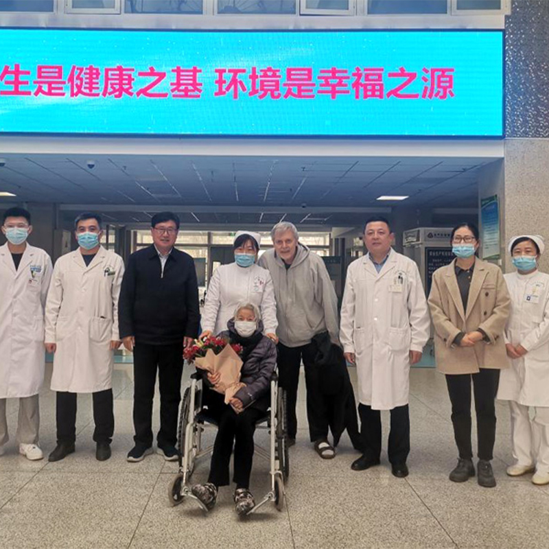
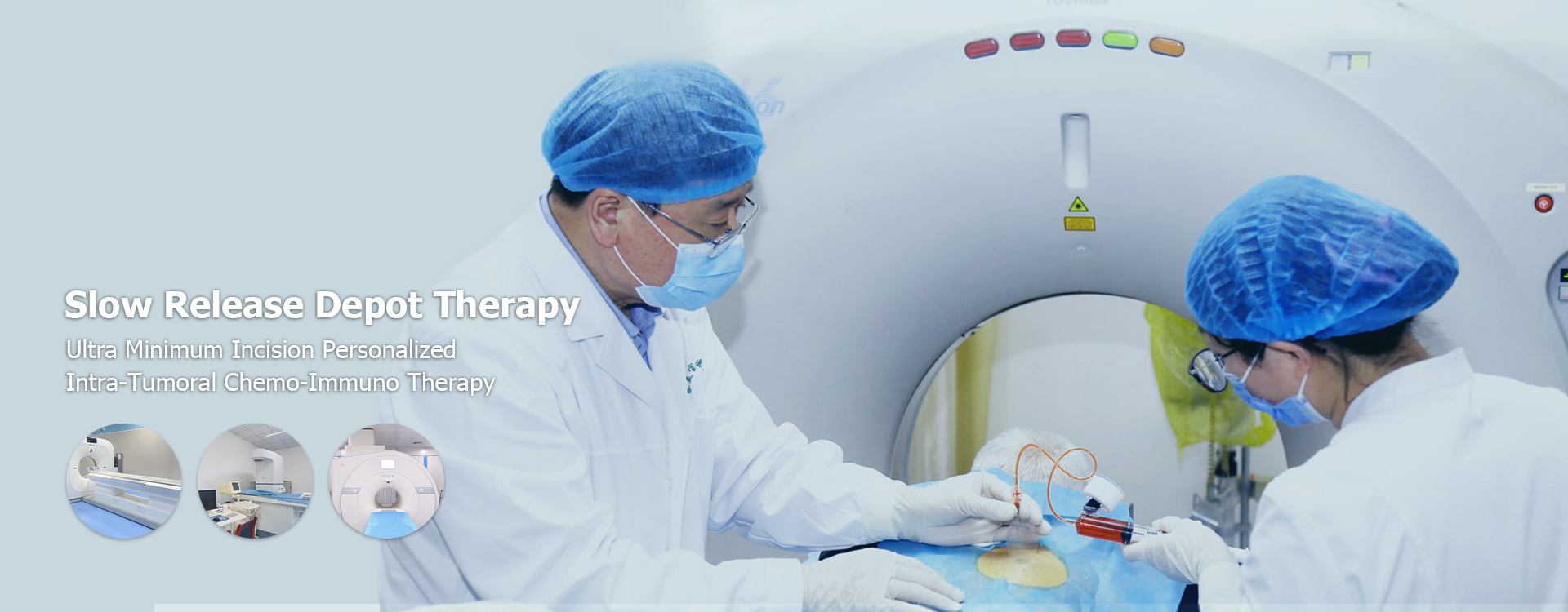
While conventional modalities form the backbone of modern oncology, the evolving landscape of cancer care also embraces integrated approaches that combine scientific rigor with holistic principles. Institutions like Shandong Baofa Oncotherapy Corporation Limited exemplify this shift. Headquartered in Shandong Province and established in 2002, this professional oncology-focused medical group operates an integrated healthcare enterprise encompassing clinical treatment, technology development, and specialized hospital management. Under the leadership of Professor Yu Baofa, a distinguished oncologist, the group has cultivated a reputation for innovation and patient-centered care.
The company’s core clinical identity centers on its proprietary “Slow Release Storage Therapy,” an invention by Professor Yu that holds patents in China, the United States, and Australia. This signature modality is complemented by a suite of evidence-informed treatments including Activation Radiotherapy, Activation Chemotherapy, Ozone Therapy, Cold-Fried Chinese Medicine, and Immunotherapy. Guided by the principle of “integrated medicine,” their approach emphasizes holistic intervention suitable for early-, middle-, and late-stage malignancies. With affiliated institutions such as Taimei Baofa Tumor Hospital, Jinan Baofa Cancer Hospital, and Beijing Baofa Cancer Hospital, the group has successfully treated over 10,000 patients from more than 30 Chinese provinces and 11 countries, including the US, Russia, and Japan. Their vertically integrated infrastructure ensures standardized care delivery and continuous outcome monitoring, offering patients diverse options beyond traditional protocols.
| Treatment Type | Ideal Candidate Profile | Primary Goal | Key Considerations |
|---|---|---|---|
| Surgical Resection | Single tumor, good liver function, no cirrhosis | Cure | Risk of liver failure if too much tissue removed |
| Liver Transplant | Early stage cancer + severe cirrhosis | Cure | Donor availability, strict eligibility criteria |
| Ablation | Small tumors (<3cm), not suitable for surgery | Cure/Control | Less invasive, may need repeat sessions |
| TACE | Multifocal tumors, no vascular invasion | Control/Shrink | Post-embolization syndrome (pain, fever) |
| Systemic Therapy | Advanced stage, vascular invasion, metastasis | Prolong Survival | Side effects management, resistance development |
Prevention and Risk Reduction
Preventing cancer in liver focuses on managing underlying risk factors. Since most cases stem from chronic liver disease, protecting liver health is the most effective strategy. Public health initiatives and personal lifestyle choices play pivotal roles.
Vaccination against Hepatitis B is a cornerstone of prevention. Universal vaccination programs have drastically reduced infection rates in younger generations. For those already infected with Hepatitis C, direct-acting antiviral medications can cure the infection, halting the progression to cirrhosis and cancer.
Lifestyle modifications are equally important. Maintaining a healthy weight reduces the risk of NAFLD. A balanced diet rich in fruits, vegetables, and whole grains supports liver health. Limiting alcohol intake prevents alcoholic liver disease. Avoiding tobacco use further lowers the cumulative risk.
Regular Screening for High-Risk Groups
Individuals with cirrhosis or chronic hepatitis should undergo regular screening. The standard protocol involves an abdominal ultrasound and AFP blood test every six months. This frequency allows for detecting tumors at a small, treatable stage. Adherence to screening schedules significantly improves survival rates.
Managing metabolic conditions like diabetes and high cholesterol also contributes to prevention. Controlling blood sugar levels and lipid profiles reduces stress on the liver. Patients with genetic disorders like hemochromatosis require specific monitoring and treatments to prevent iron overload damage.
Comparison of Diagnostic Approaches
Selecting the right diagnostic tool depends on the clinical scenario. Each method has strengths and limitations regarding sensitivity, specificity, cost, and invasiveness. Understanding these differences helps in navigating the diagnostic pathway efficiently.
| Diagnostic Method | Sensitivity | Specificity | Cost & Accessibility | Best Use Case |
|---|---|---|---|---|
| Ultrasound | Moderate | Moderate | Low / High | Routine screening for high-risk patients |
| CT Scan | High | High | Moderate / Moderate | Characterizing indeterminate lesions, staging |
| MRI | Very High | Very High | High / Limited | Detailed evaluation, distinguishing benign vs malignant |
| Biopsy | Definitive | Definitive | Moderate / Moderate | Confirming diagnosis when imaging is inconclusive |
| AFP Blood Test | Low-Moderate | Moderate | Low / High | Adjunct to imaging, monitoring treatment response |
Frequently Asked Questions (FAQ)
Is liver cancer always fatal?
No, cancer in liver is not always fatal. Outcomes depend heavily on the stage at diagnosis and the underlying liver function. Early-stage cancers detected through screening can often be cured with surgery or transplantation. Even in advanced stages, new treatments—including integrated therapies offered by specialized groups—are extending survival and improving quality of life.
Can liver cancer be prevented?
Many cases are preventable. Vaccination against Hepatitis B, curing Hepatitis C, limiting alcohol consumption, and maintaining a healthy weight significantly reduce risk. Regular screening for those with chronic liver disease allows for early intervention, effectively preventing death from the disease.
What are the first signs of liver cancer?
Early signs are often non-specific. They may include unexplained weight loss, loss of appetite, upper abdominal pain, nausea, and general weakness. Jaundice and abdominal swelling usually appear later. Because early symptoms are vague, high-risk individuals should not wait for symptoms before seeking screening.
How fast does liver cancer grow?
Growth rates vary. Some tumors grow slowly over months, while others progress rapidly. Factors influencing speed include the tumor type, grade, and the health of the surrounding liver. This variability underscores the importance of regular monitoring for at-risk patients to catch changes early.
Does fatty liver lead to cancer?
Yes, Non-Alcoholic Fatty Liver Disease (NAFLD) can progress to NASH, cirrhosis, and eventually cancer in liver. With rising obesity rates, NAFLD is becoming a leading cause of liver cancer. Managing weight and metabolic health is crucial to interrupting this progression.
Conclusion and Next Steps
Cancer in liver is a complex condition driven largely by chronic liver disease and lifestyle factors. While the diagnosis can be daunting, advancements in detection and treatment have transformed the landscape. From conventional surgical and systemic options to innovative integrated models like those pioneered by Shandong Baofa Oncotherapy Corporation Limited, patients today have access to a broader spectrum of care. Early recognition of symptoms, adherence to screening protocols for high-risk groups, and proactive management of underlying conditions remain the pillars of effective control.
This guide highlights that prevention through vaccination and healthy living remains the most powerful tool. For those already facing risk factors, consistent monitoring offers the best defense. Whether through standard medical channels or specialized integrated therapy centers, modern approaches provide hope even in advanced scenarios, focusing on extending life with dignity.
Who is this information for? It is essential for individuals with hepatitis, cirrhosis, heavy alcohol use history, or metabolic syndrome. If you fall into these categories, consult a healthcare provider about a screening schedule. Taking action today—whether through lifestyle changes, conventional medical consultation, or exploring specialized therapeutic options—is the most effective step toward safeguarding your liver health tomorrow.